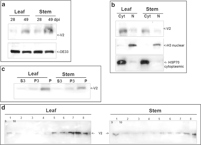
Figure 1.
Detection of TYLCV V2 in tomato leaf and stem 28 and 49 days (dpi) after infection. a: western blot of V2 and OE33 (chloroplast protein used as an internal marker). b: western blot of V2 in leaf and stem tissues at 49 dpi fractioned into cytoplasmic/membrane (Cyt) and nuclear (N) components; cytoplasmic Hsp70 and nuclear histone H3 used as internal markers to assess the fraction purity. c: western blot of V2 in leaf and stem tissues of 49 dpi fractionated into insoluble debris and cell wall (P), 3000 g pellet (P3) and soluble protein (S3). d: western blot analysis of V2, distributed in linear 10-50% sucrose gradients from leaf and stem native protein extracts at 49 dpi; gradients were divided into 10 fractions, 1 (top) to 10 (bottom) and aliquots were subjected to SDS-PAGE.

