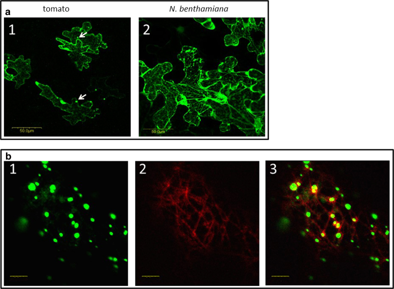
Figure 4.
Expression of fusion V2:GFP in epidermal cells of tomato and N. benthamiana leaves. a: tomato (1) and N. benthamiana (2) leaves infiltrated with A. tumefaciens carrying the V2:GFP expression plasmid. Bar: 50 μm. b: N. benthamiana leaves infiltrated with A. tumefaciens carrying the V2:GFP and MAP4:RFP expression plasmids: 1. V2:GFP (green); 2. microtubule marker MAP4:RFP (red); 3. merge of V2:GFP and MAP4:RFP, V2 co-localization with microtubules is indicted in yellow. Bar: 5 μm.

