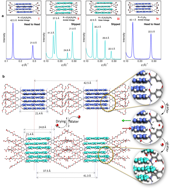
Figure 5. Mechanism of the fluorescence change based on molecular packing.
(a) SAXS pattern of PE1 in the (i) absence and (ii) presence of water, (iii) PE2 and (iv) PE3. The corresponding molecular arrangements are shown on the top of the SAXS patterns. (b) Schematic illustration of sliding of the PE1 molecule in the absence and presence of water on paper surface. Disruption of H-bonds and the breathing of the oxyethylene chains in presence of water experience an inward pushing of the molecules resulting in the change of an H-type (B-phase) to J-type (C-phase) packing. The images in panel ‘b’ (right) show the zoomed portion of the molecular arrangement illustrating the H-bond breaking and molecular sliding (arrows show the direction of sliding).

