Figure 3.

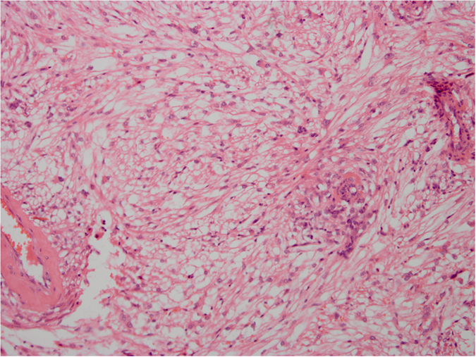
HE staining. The epithelioid cells are not distributed uniformly. Atypical epithelioid cells with abundant cytoplasm, vesicular nuclei, prominent nucleoli, and large nuclear size can be seen. Original magnification ×200.

HE staining. The epithelioid cells are not distributed uniformly. Atypical epithelioid cells with abundant cytoplasm, vesicular nuclei, prominent nucleoli, and large nuclear size can be seen. Original magnification ×200.