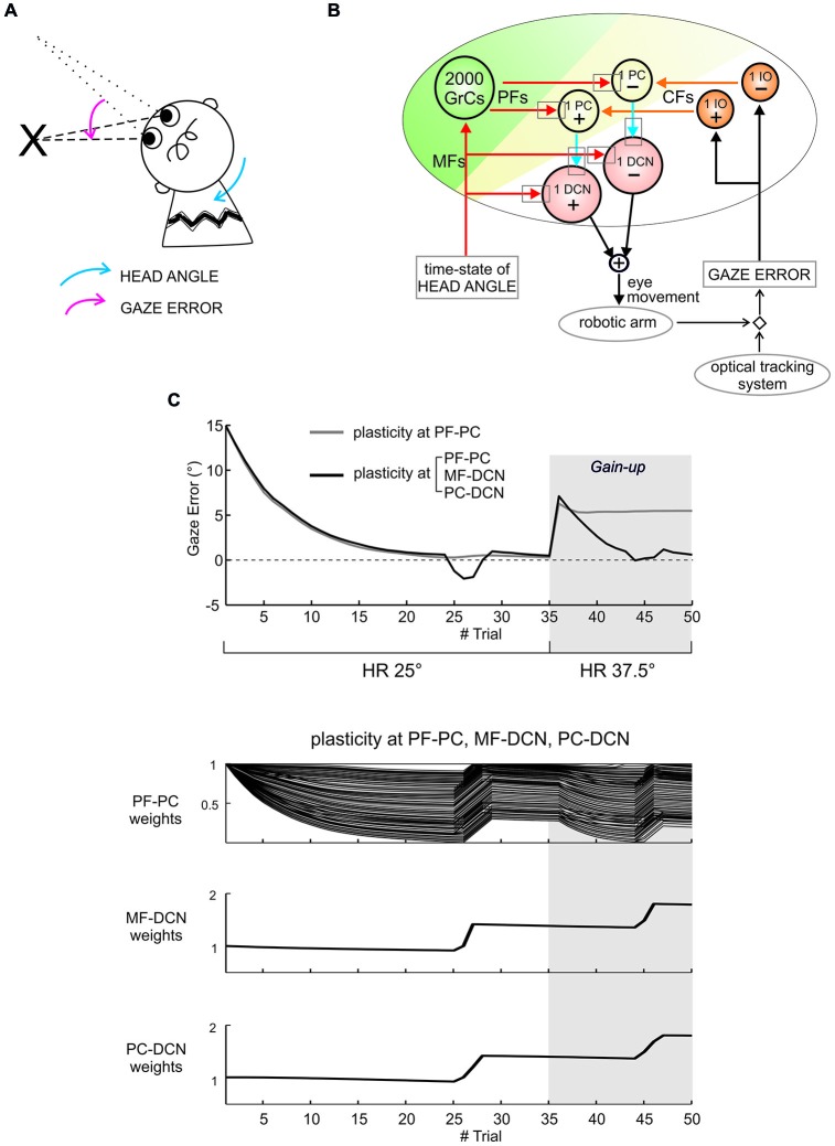
Figure 3.
Distributed cerebellar plasticity in real-robot sensorimotor vestibulo-ocular reflex (VOR) task. (A) Human-like VOR task: the arrows show the head angle rotation (blue arrow) and the angle of gaze error (pink arrow). (B) Cerebellar model with VOR-specific input and output signals. The plasticity sites are indicated by the gray rectangles (all plasticities are bidirectional with LTP and LTD rules defined and calibrated according to experimental observations, see (Casellato et al., 2014, 2015; Luque et al., 2014)). Red and orange arrows indicate excitatory connections from mossy fibers (MFs) and CFs, respectively. Blue arrows indicate inhibitory connections. The head vestibular stimulus represents the system time-state, decoded by the granular layer. The gaze error is fed into the CF pathway, and the DCN neurons modulate compensatory eye movements. (C) Gain-up VOR test: after 35 trials, the head rotation (HR) was increased 1.5 times (from 25° to 37.5°), and imposed for other 15 trials. The curves report the gaze error within each of the total 50 trials, implementing plasticity at a single site (PF-PC connection, in gray) or at multiple sites (PF-PC, MF-DCN and PC-DCN connections, in black). With one or three plasticities, the robot compensated equally well for HR. However, while plasticity at the PF-PC connection alone proved unable to change the gain and to correct for the increased HR, combined plasticities at PF-PC, MF-DCN and PC-DCN were able to rescale the response and adapt to the new HR angle. The three bottom plots show synaptic weights at the end of each trial for the three synapses involved, referring to the case of plasticity at the three connections. Indeed, the transfer from cortical to nuclear sites made the PF-PC synapses ready for further plasticity, making them able to react to perturbations suddenly presented to the system. Modified from Casellato et al. (2015).

