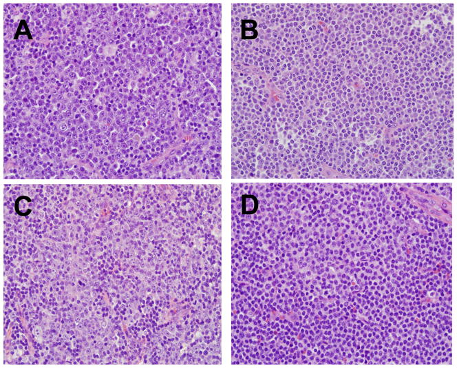
Figure 1. Lymph node Histology in CLL.
Proliferation centers characterized by paraimmunoblasts (prolymphocytes) are a characteristic feature of lymph nodes involved by CLL. Prominent proliferation centers with >40% paraimmunoblasts are shown in sections A to C. In section B there is a more subtle increase in paraimmunoblasts interspersed with small CLL lymphocytes. D: Example of a more typical appearance of CLL with a predominance of small lymphocytes and foci of proliferation center formation. (H&E stain, Nikon objective 20x lens with 200x magnification, Olympus DP controller and Nikon Eclipse 80i camera)

