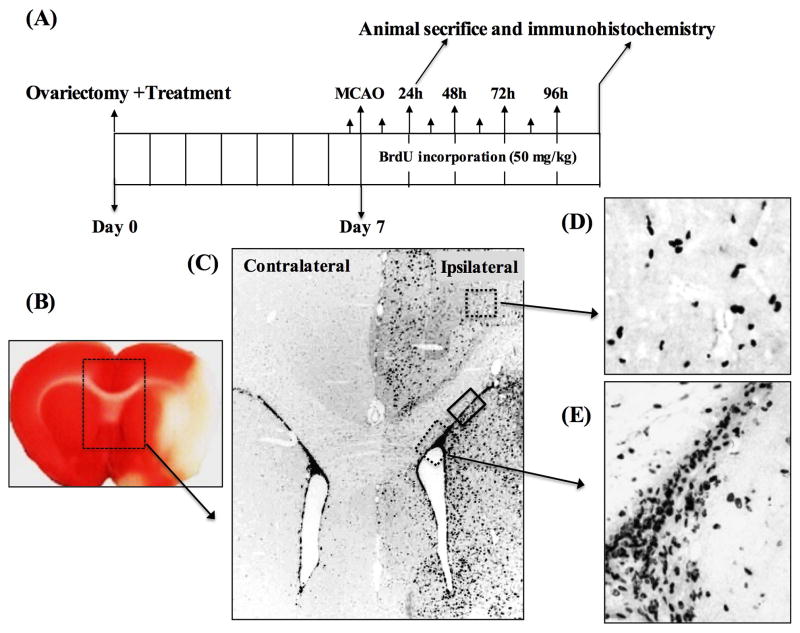
Figure 1. Ischemia induces neurogenesis in the SVZ of ovariectomized female rats.
A) Shows the experimental design for the neurogenesis experiments. Animals were ovariectomized and E2 or SERM treatment initiated immediately. MCAO was performed one week later. BrdU (50 mg/kg, ip) was injected 15 min prior to MCAO followed by two BrdU injections daily for 96 h post MCAO. Animals were sacrificed at day-1 and day-5 post MCAO and processed for immunohistochemistry. B), a TTC stained coronal section of the brain showing the development of infarct at 24h (day-1) post MCAO. The box region illustrates the contralateral and ipsilateral lateral ventricles (SVZ), which was magnified to see the pattern of NPCs proliferation in response to ischemia. C) Shows the DAB staining pattern of BrdU+ cells in the contralateral and ipsilateral SVZ from a placebo-treated animal at day-5 post MCAO. The box regions depict the areas of ipsilateral cortex (upper dashed box) and the ipsilateral ventral SVZ (lower dotted box) and the dorsal SVZ (solid box) of the ipsilateral hemisphere showing the migration of BrdU+ cells following MCAO. D and E) show the larger views of DAB staining pattern of BrdU+ cells detected in ipsilateral cortex and ventral SVZ regions, respectively.

