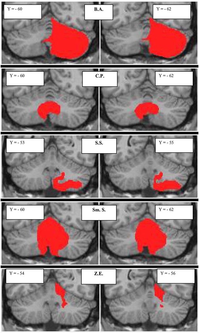
Figure 1.

Subjects with focal cerebellar lesions. Lesion extensions were assessed on 3D-T1-MPRAGEs after spatial normalization and overlaid onto a coronal T1-weighted template from Schmahmann et al. (2000). For each subject, the lesion is shown in two representative coronal sections. The case code is as in Table 2.
