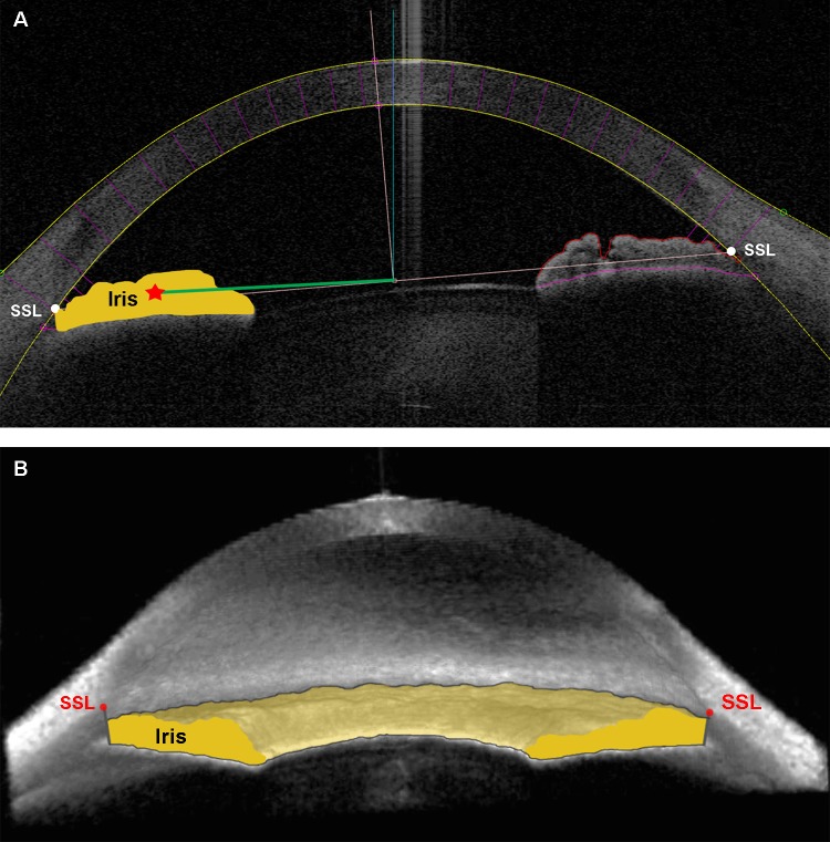
Figure 2.
Iris volume. (A) The 2D and (B) 3D ASOCT angle images exhibiting borders for IV calculation. Iris cross-sectional area is the area bordered by anterior iris surface, anterior surface of the demarcation line created by the reflection of the posterior layer of pigment epithelium, and the line drawn from SSL and parallel to visual axis. RICSA is represented by the green line. (A). Iris volume is the integrated iris cross-sectional area measured around the angle (B). The SSL appears above the iris plane in the 3D view of this open-angle eye.15

