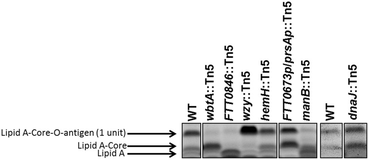
Figure 4.
Lipopolysaccharide core structures of mutant strains. An SDS-PAGE gel was loaded with ~14 μg of dried material per mutant per well. The gel was stained with Emerald green, and is labeled to indicate lipid A—core—O-antigen (one unit), lipid A—core (no O-antigen subunit), or free lipid A (indicated by black arrows).

