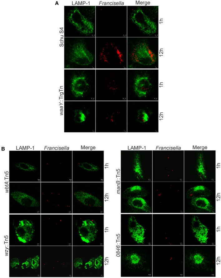
Figure 6.
Microscopy of infected MDMs with wbtA, wzy, manB, and FTT0846 mutants. Confocal images show Lamp-1 in green and bacteria in red. MDMs were infected with (A) wild type Schu S4 as the positive control, waaY::TrgTn as a negative control, and with (B) wbtA::Tn5, wzy::Tn5, manB::Tn5, or FTT0846::Tn5 strains at an MOI of 100:1. Samples were fixed and processed at 1 and 12 h post infection.

