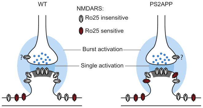
Fig. 6.

Model of aberrant GluN2B function in PS2APP mice. Glutamate from single synaptic activations (dark blue) only reaches synaptic NMDARs while bursts of synaptic activation lead to glutamate reaching perisynaptic pools of NMDARs (light blue). In wt mice, these perisynaptic NMDARs are generally insensitive to Ro25 (white), while in PS2APP mice, these peri-synaptic NMDARs are more sensitive to Ro25 (red). Extrasynaptic NMDARs located farther from the synapse are distinct from the other pools. Question marks indicate the unknown Ro25 sensitivity of presynaptic NMDARs (gray). While an altered physical presence of GluN2B-NMDARs is depicted, the observed functional phenotypes could also result from altered activation caused by impaired glutamate transport and greater spread of glutamate from the synapses during burst activation.
