
Fig. 5.
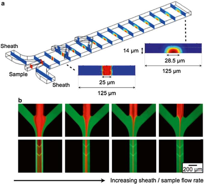
To enhance the μHall signal, a hydrodynamic focusing structure was used to bring individual cells close to the μHall sensors. Cells entering the chip though the sample input are pushed towards the center of the channel by the lateral sheaths and are pushed vertically to the bottom of the channel by the chevron patterns. (a) The results of a finite element computer simulation. The sheath fluid is colored blue and the sample fluid in colored red. (b) Fluorescence micrographs, demonstrating flow focusing. The sheath fluid was labeled with fluorescein and the sample was labeled with rhodamine. As the sheath/sample flow rate ratio increased, the sample flow was focused to an increasingly narrow region in the center of the chip