
Fig. 7.
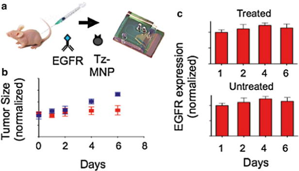
Molecular analysis of fine needle aspirates on a μHall chip. (a) The μHall chip was used to profile EGFR expression of cells obtained by fine needle aspirate from mice with a xenografted tumor. Mice bearing xenografted tumors were treated with geldanamycin for 6 days or left untreated (n = 6 per group). (b) Rate of tumor growth in untreated mice and mice treated with geldanamycin. The blue and red data points represent untreated and treated mice, respectively. (c) Tumor samples were screened by the μHall chip to monitor the changes in EGFR expression over the course of the drug treatment (Color figure online)