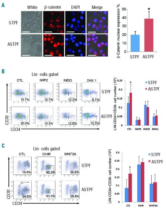
Figure 6.

Wnt signaling participated in ANGPTL7-stimulation in hematopoietic stem and progenitor cells (HSPCs). (A) (Left) Immunofluorescence analysis of subcellular localization of β-catenin (red) in CD34+ human HSPCs treated with ANGPTL7 (ASTPF) or without ANGPTL7 (STPF) for 24 h. Scale bar: 100 μm. Nuclei were stained with DAPI (blue). In merged magnification images, the overlap of blue and red indicated the localization of β-catenin in nuclei. (Right) Chart depicting the percentage of β-catenin nuclear expression in CD34+ human HSPCs treated as indicated. Error bars show +/− s.e.m. of triplicates from three independent experiments; *P≤0.05 for bar 1 versus bar 2. (B) (Left) Representative FACS profiles of CD34+ purified human umbilical cord blood nucleated cells (hUCBNCs) cultured in ASTPF and STPF conditions with or without Wnt inhibitors (IWP2, INDO, or DKK1). (Right) Summary of absolute numbers of Lin-CD34+CD38− cells in purified CD34+ hUCBNCs cultured in ASTPF and STPF conditions with or without Wnt inhibitors (IWP2, INDO, or DKK1). Data represent mean+s.e.m. (n=3). *P≤0.05 for bar 1 versus bar 2. (C) (Left) Representative FACS profiles of CD34+ purified hUCBNCs cultured in ASTPF and STPF conditions with or without Wnt activators (CHIR or WNT3A). (Right) Summary of absolute numbers of Lin-CD34+CD38− cells in purified CD34+ hUCBNCs cultured in ASTPF and STPF conditions with or without Wnt activators (CHIR or WNT3A). Data represent mean+s.e.m. (n=3); *P≤0.05 versus bar 2 for bar 1.
