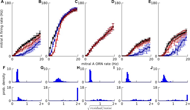
Fig 6. . Factors affecting linearity.
A-E: Mitral input-output curves simulated in a default network (Figs 2D and 4D(i)) and modifications thereof. In each case mitral firing rate is plotted against ORN firing rate to the tuft of mitral cell A. Activity-dependent lateral inhibition to A, mediated by granule cells, is assessed in each case by considering a lateral ‘super-inhibiting’ mitral cell B receiving Poisson ORN input at 0 Hz (black squares), 5 Hz (red diamonds) and 10 Hz (blue circles). A. default. B. PG cells removed. C. granule cells removed. D, E: The default network is modified to have stronger PG cell excitation yielding half-Mexican-hat profiles i.e. suppression of mitral firing for low ORN input. D. ORN→PG and mitral→PG synaptic strengths are increased by 2.4 times for both plateau-ing and low threshold spiking PG cells. E. ORN→PG and mitral→PG synaptic strengths are increased by 6 times for plateau-ing PG cells but unchanged for low threshold spiking PG cells. F-J. The histograms of for fits to single odor random pulse-trains (top row) and for predictions to two-odor random pulse-trains (bottom row) corresponding to above A-E cases respectively, from 30 network instances, each with different two odors. All values for are in the right-most bin. The mitral input-output curve saturated to a small extent on removing granule cells (C) causing a worsening of the linear fits and predictions. But on removing PG cells (B), the saturation was much stronger and the lateral inhibition for 10 Hz input to lateral mitral cell B was less than that for 5 Hz. On modifying the input-output curve to have an initial supra-linear region (half-Mexican-hat) (D,E), the predictions were often seen to be supra-linear compared to the fits. Also, the lateral inhibition turned on with a threshold, i.e. negligible for 5 Hz input to mitral cell B but large for 10 Hz, since the lateral mitral cell B had the same non-linear input-output curve.

