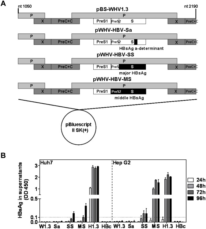
Fig 2. Constructions of recombinant WHV-HBV genomes and transient transfection of recombinant WHV-HBV genomes in hepatoma cells.
(A) The schematic map of recombinant WHV-HBV genomes. pBS-WHV1.3 contained a 1.3 fold overlength WHV genome in pBluescript II SK(+) vector and was used as backbone. The respective WHV genome regions were replaced by the corresponding HBV sequences, shown as black bars. The plasmids pWHV-HBV-Sa, pWHV-HBV-SS, and pWHV-HBV-MS contained HBV sequences encoding HBsAg a-determinant only, major HBsAg and middle HBsAg, respectively. (B) HBsAg expression in the supernatant of the transfected hepatoma cells. Huh7 and HepG2 cells were transiently transfected with plasmids of pBS-WHV1.3 (W1.3), pWHV-HBV-Sa (Sa), pWHV-HBV-SS (SS), pWHV-HBV-MS (MS), pBS-HBV1.3 (H1.3), and pHBc (HBc). The culture supernatants were collected at 24, 48, 72, and 96 hours after transfection for the detection of HBsAg by ELISA. The results were read at OD 450 nm. The cut off value was set as 0.1 and indicated by the dotted line.

