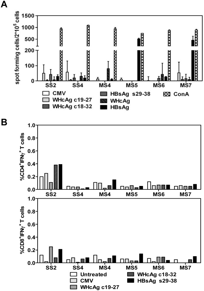
Fig 7. The specific T cell responses to WHcAg and HBsAg in BALB/c mice received HI with the recombinant WHV-HBV genomes.
Mouse splenocytes were taken at week 36 after HI with pWHV-HBV-SS (SS) and pWHV-HBV-MS (MS). The splenocytes were cultured in presence of HBsAg-derived peptide s29-38, WHcAg-derived peptides c19-27 and c18-32, WHcAg, and HBsAg. (A) IFN-γ producing cells were detected by ELISpot assay. The results were shown as spot-forming cells per 2×10⁵ cells. ConA and an unrelated CMV peptide were used as positive and negative control, respectively. (B) The frequencies (%) of IFN-γ producing CD4+ and CD8+ T cells in mouse splenocytes were analyzed by staining of cell surface markers and intracellular IFN-γ and flow cytometry. Untreated: splenocytes cultured without specific peptides.

