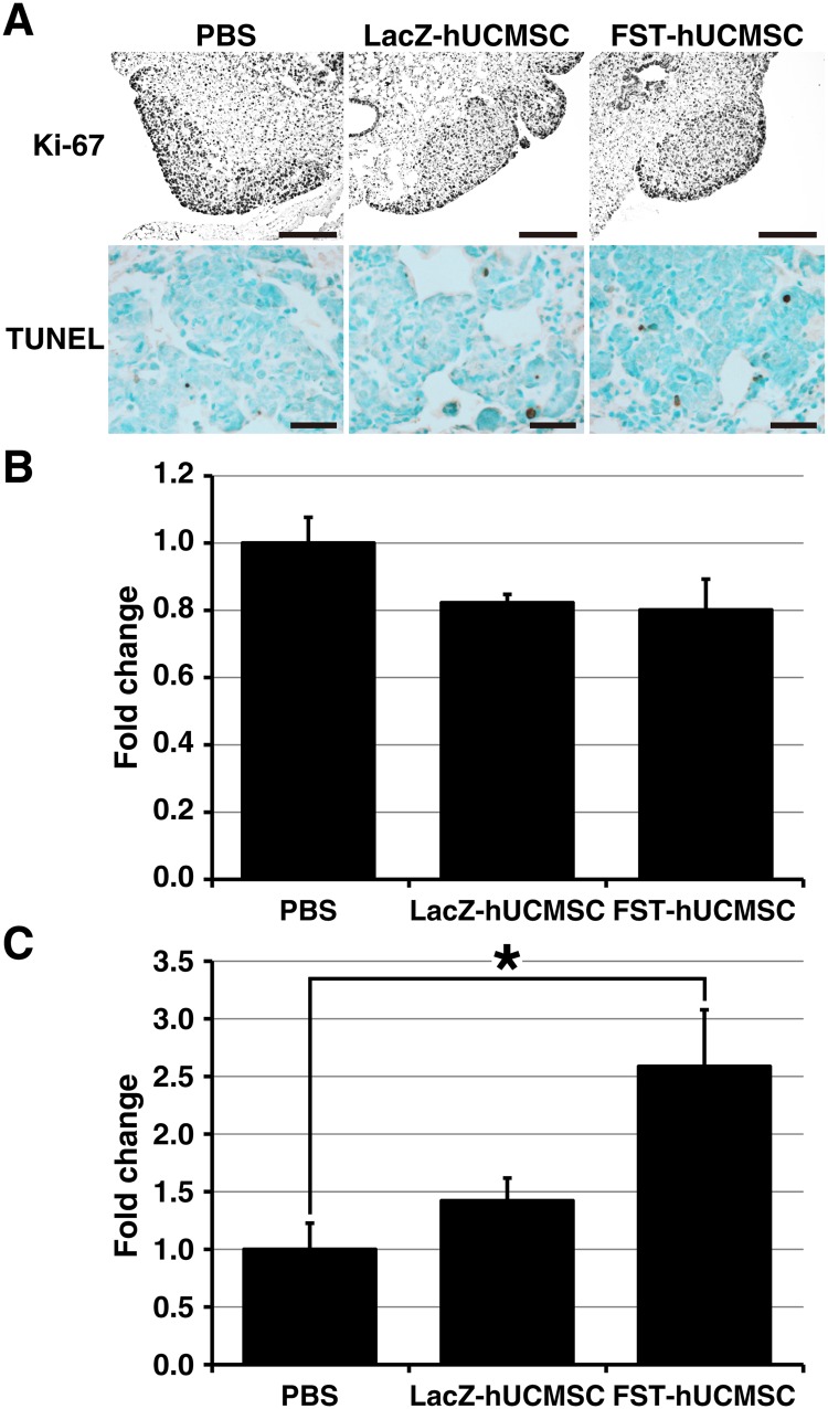
Fig 5. Immunohistochemical analysis of cell proliferation (A and B) and apoptosis (A and C) in MDA-231 graft tumors in SCID mouse lungs treated with either PBS, LacZ- or FST-hUCMSC.
A, microscopic images of immunohistochemistry for Ki-67 (top 3 panels) at 20x and TUNEL assay (bottom 3 panels) at 40x. B, treatment with LacZ- or FST-hUCMSC had no significant effect on proliferation of the tumor cells. C, the TUNEL positive cells were significantly increased in the tumors of mice treated with FST-hUCMSC. * p < 0.05 as compared to the level of the PBS-treated control.

