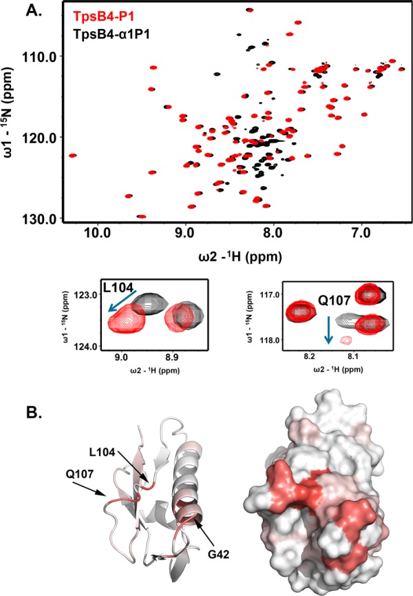
Figure 3.

The TpsB4 N-terminal helix linker interacts with its POTRA1 domain. (A) Comparison of 1H-15N HSQC NMR spectra for 15N-labelled TpsB4-P1 (red) and TpsB4α1P1 (black), with chemical shift perturbations associated with L104 and Q107 in the β4-β5 loop expanded. (B) Chemical shift mapping on TpsB4-P1 of peak perturbation between 1H-15N HSQC NMR spectra of 15N-labelled TpsB4-P1 and TpsB4-α1P1. Deeper red areas indicate residues in closer vicinity to the TpsB4-α1P1 N-terminal plug helix/linker. Numbering is based on mature TpsB4 minus the N-terminal signal sequence. An interactive view is available in the electronic version of the article
