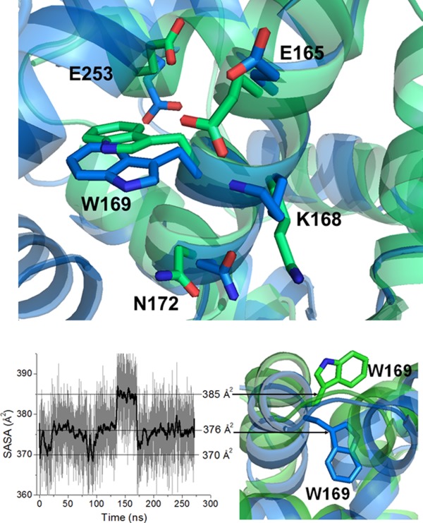
Figure 7.

Top: charged and polar amino acid residues surrounding Trp169 in the structure of DREAM (PBD entry 2JUL, shown in blue) and C-terminal domain of DREAM (PBD entry 2E6W, shown in green). Bottom: Left: SASA of Trp169 in DREAM during 270 ns of the MD trajectory. Right: Partially buried (in blue) and solvent exposed (in green) orientation of Trp169 side chain in DREAM structure.
