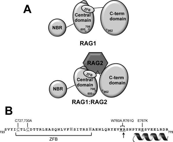
Figure 6.

A: Model of the RAG1-RAG2 complex. The upper schematic shows the three domains in core RAG1 (NBD, central, and C-terminal domains). The ZFB is denoted as an oval, and the three active site residues (D600, D708, and E962) labeled and indicated with asterisks. The pentagon adjacent to the ZFB represents a region of charged residues in the central domain that upon mutation were previously shown to disrupt complex formation with RAG2.18,27 For simplicity, only a monomer of core RAG1 is represented. The lower schematic depicts RAG2 bridging the central and C-terminal domains, resulting in a closer juxtaposition of D600 and D708 with E962. B: The sequence of RAG1 that showed decreased susceptibility to proteases in the presence of RAG2. The border between the central and C-terminal domains is marked with an arrow. The sequence of the putative ZFB is bracketed, and likely Cys and His zinc-coordinating residues are denoted in a larger gray font. The helix denotes the region at the C-terminal end of this peptide predicted to be alpha helical. The core RAG1 mutants are indicated above the sequence, and the corresponding residues within the sequence that were mutated are underlined.
