Figure 1.
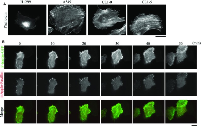
Staining for F-actin in non-small cell lung adenocarcinoma cells reveals that the cells isolated from a secondary cancer of the lymph nodes, namely H1299 cells, do not form bundled actin filaments (stress fibres) during cell migration. (A) CL1-0, CL1-5, A549 and H1299 cells were immunostained with fluorescent phalloidin, to localize F-actin; bar 15 μm. (B) Time-lapse TIRF images of H1299 cells expressing F-tractin-GFP and mApple-paxillin showing the dynamics of F-actin and FAs, respectively; bar 15 μm.

