Figure 3.
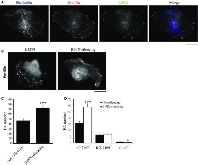
β-PIX is mainly localized at FAs and regulates FA dynamics. (A) H1299 cells were immunostained to localize F-actin (blue; phalloidin), paxillin (red; as a focal adhesion marker), and β-PIX (green); bar 15 μm. (B–D) Non-silencing and β-PIX-silencing H1299 cells were immunostained with paxillin, to localize FAs, and imaged by TIRF microscopy (B); bar 15 μm. The number (C) and size distribution (D) of segmented paxillin-marked adhesions within the cells. Data are mean ± SEM (non-silencing, n = 1118 FAs/24 cells; β-PIX-silencing, n = 1744 FAs/24 cells). ***P < 0.001, *P < 0.05, in comparison with non-silencing cells.

