Figure 5.

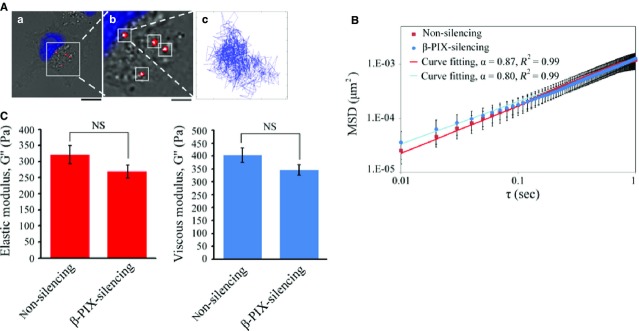
Multiple-particle tracking in non-silencing and β-PIX-silencing H1299 cells. (A) Typical trajectories of 100 nm diameter fluorescent carboxylated polystyrene beads (red) embedded in the cytoplasm of H1299 cells with the nucleus was labelled in blue (a and b). Beads were delivered into the cells; and after overnight incubation, the two-dimensional Brownian motion of the intracellular fluorescent beads was tracked and recorded at 10 msec. temporal resolution and 20 nm spatial resolution for 10 sec. (a) Bar 15 μm; (b) bar 5 μm. (B) The mean squared displacement (MSDs) as a function of the time lag (τ) of the imbedded fluorescent particles in non-silencing and β-PIX-silencing H1299 cells. The MSDs were fit to the power law <Δr2(τ)> = Aτα. Data are mean ± SEM (non-silencing, n = 201 beads/35 cells; β-PIX-silencing, n = 513 beads/45 cells). (C) Intracellular elastic modulus (G′) and viscous modulus (G″) of non-silencing and β-PIX-silencing H1299 cells at 10 Hz (non-silencing, n = 201 beads/35 cells; β-PIX-silencing, n = 513 beads/45 cells). Data are mean ± SEM. NS, no significance.
