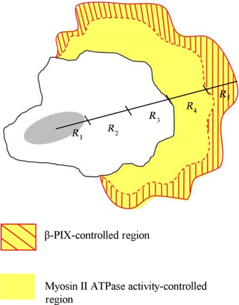
Figure 8.

A model showing β-PIX-mediated signalling and myosin II ATPase-mediated signalling spatially controlling intracellular viscoelasticity and actin cytoskeleton organization, both of which contribute to the coordination of the cell migration process in non-small cell lung adenocarcinoma cells. In a highly metastatic lung cancer cell isolated from a secondary lung cancer of the lymph nodes, namely H1299 cells, the β-PIX-mediated signals are fully activated, as the cells lack bundled actin filaments (stress fibres). β-PIX mainly is localized at the immature FAs that drive lamellipodia extension, enhancing the integrity of the actin networks, up-regulating intracellular viscoelasticity at the cell periphery (R5 region), and control cell polarity, which then promotes directed cell migration. When compared to the cells with stress fibres, our findings show that there are a variety of different functions for myosin II ATPase, including promoting the organization of the actin cytoskeleton, changing intracellular viscoelasticity in the R4 and R5 regions, and modulating cell migration. For details, see Discussion.
