
Figure 4.
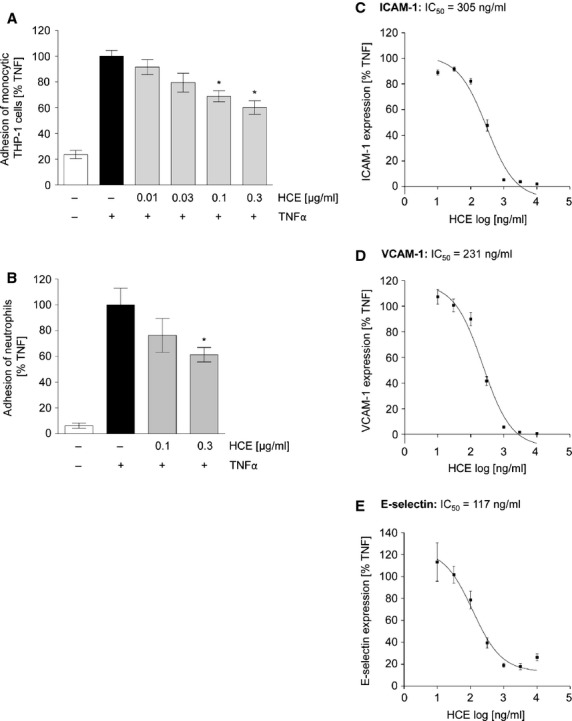
HCE strongly reduces leucocyte adhesion by blocking the expression of endothelial adhesion molecules. (A) After HCE pre-treatment (10–300 ng/ml for 30 min.) HMECs were treated with TNFα (10 ng/ml) for 6 hrs. Cell-Tracker™ Green-labelled THP-1 monocytic cells were added (3 × 10⁵ cells per well) and were allowed to adhere for 1 hr. The fluorescence of adhered monocytic THP-1 cells was measured (ex: 485 nm; em: 535 nm). N = 4; *P ≤ 0.05, versusTNFα. (B) Confluent primary endothelial cells were left untreated or were pre-incubated with HCE (0.1 and 0.3 μg/ml) for 30 min. Then they were treated with TNFα (10 ng/ml) for 24 hrs. Human neutrophil granulocytes were added (105 cells per well) and were allowed to adhere for 45 min. To determine the amount of adhered neutrophils the activity of myeloperoxidase was measured. N = 3; *P ≤ 0.05 versusTNFα. (C and D) Adhesion molecule expression on the endothelial cell surface was determined by flow cytometry. HUVECs were either left untreated or were pre-incubated with HCE (10 ng/ml–10 μg/ml) for 30 min. Afterwards, cells were treated with TNFα (10 ng/ml) for 24 hrs (C, ICAM-1; D, VCAM-1) or for 6 hrs (E, E-selectin). N = 3.