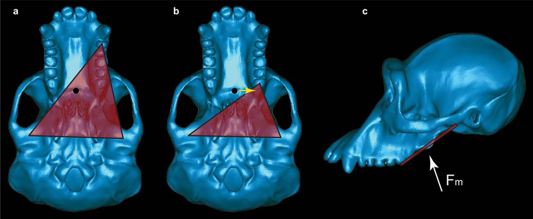
Fig. 1.
The constrained lever model. A: Basal view of a chimpanzee skull illustrating the triangle of support (red) during a bite on a mesially positioned tooth. The resultant of the masticatory muscle forces is indicated by the black circle, and for simplicity is assumed in this case to be directed perpendicular to the plane of the image (but see below). The exact location of the muscle resultant cannot be known with certainty, but will be found in the midline if the muscles on the right and left sides of the body are acting with equal activity levels. Moreover, the resultant cannot be found anterior to the distal most teeth (in this case, the third molars). Thus, the resultant shown here is in its anterior-most position under the assumption that the adductor muscles are acting with bilateral symmetry. Note that the resultant in this bite falls within the triangle of support (red triangle) defined by the bite point and the two TMJs. B: A bite on a more distally placed tooth. The muscle resultant is now found outside the triangle. By reducing the balancing-side muscle force, it is possible to shift the position of the resultant toward the working-side (yellow arrow) so that the resultant is once again within the triangle. Note that if the resultant were to be found anterior to the bite point, it would be impossible to shift the resultant into the triangle. C: Lateral view of the masticatory apparatus. Note that the triangle of support (red line) is not horizontal, and that the muscle resultant (Fm, white arrow) may not be oriented perpendicular to the triangle, nor positioned in the same plane as the distal-most teeth.

