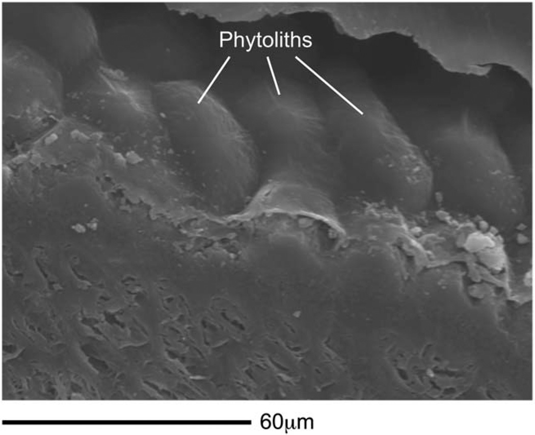
Fig. 9.
Phytoliths on seed. Scanning electron micrograph of the pericarp of a sedge seed, Cyperus bulbosus. Energy dispersive spectroscopy reveals that the rounded, pod-like structures on the cell wall of the pericarp are densely packed phytoliths (Supporting Information Fig. A2). The flat, sectioned face of the seed is below the phytoliths.

