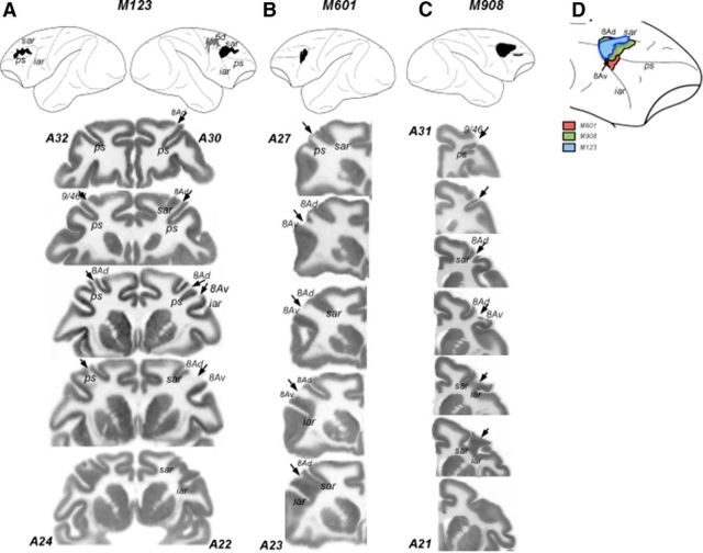
Figure 2.
Lesion reconstructions. Top, Damage from both surgical lesions and unintended damage are shown for all three animals; these have been reconstructed from histological sections and projected onto a lateral view of the macaque brain. Both hemispheres are shown for M123 (A); only the hemispheres with damage are shown for M601 (B) and M908 (C). Black areas indicate areas of damage to LPFC, and gray areas indicate damage to the dorsal premotor cortex. Numbers correspond to anteroposterior (AP) distance (mm) from the interaural plane. The lines delineate the region represented in the coronal sections shown below. Bottom, Coronal sections/slices showing damage. A–C, Cytochrome oxidase stained histological sections showing the damage in in the three animals. Discontinuities in the gray matter are the damaged regions devoid of neurons and indicated by the arrows. Numbers correspond to AP distance from the interaural plane. For M123, the two hemispheres shown in a single section are in slightly different AP coordinates. The estimates of AP coordinates are based on the Paxinos et al. (2000) stereotaxic atlas. D, Comparison of lesions in the three monkeys shown on the lateral view of the right hemisphere. The lesions were transferred onto the same hemisphere to visualize the extent of overlap between the damage in individual animals. The diagram only shows the lesions in the prearcuate region. ps, Principal sulcus; sar, superior arcuate; iar, inferior arcuate.

