
Figure 4.
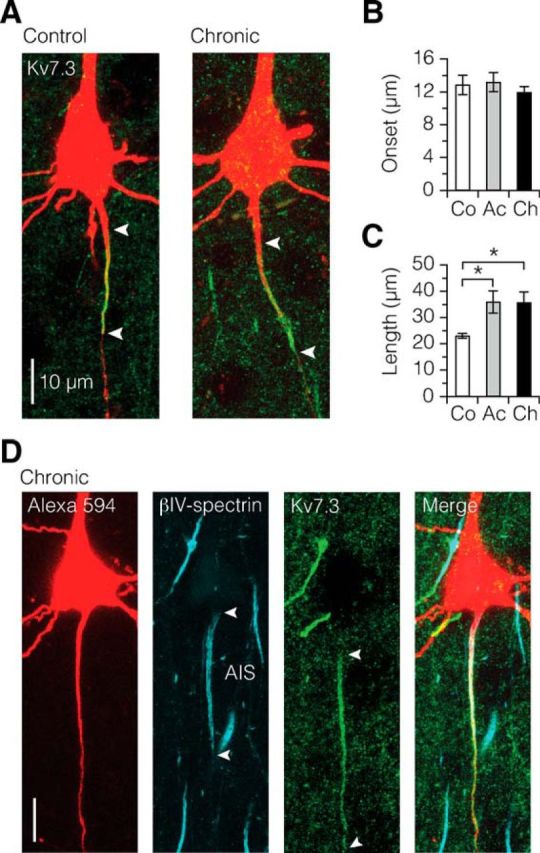
Kv7.3 ion channel expression continues into the first internode in demyelination. A–C, Kv7.3 immunofluorescence labeling (green) reveals a more extensive distribution of signal in demyelinated axons. Length: control versus acute, *p = 0.0375; control versus chronic, *p = 0.0396. Control, n = 8; acute, n = 10; chronic, n = 16. Data are presented as mean ± SEM. D, Confocal z projection of triple immunolabeling of a chronically demyelinated L5 neuron, showing the diffuse expression of Kv7.3 into the formerly myelinated first internode independently of βIV-spectrin coexpression. This diffuse expression of Kv7.3 was found in both acute (n = 3 neurons) and chronic demyelinated (n = 4 neurons) group with expression length range of 44–82 μm.