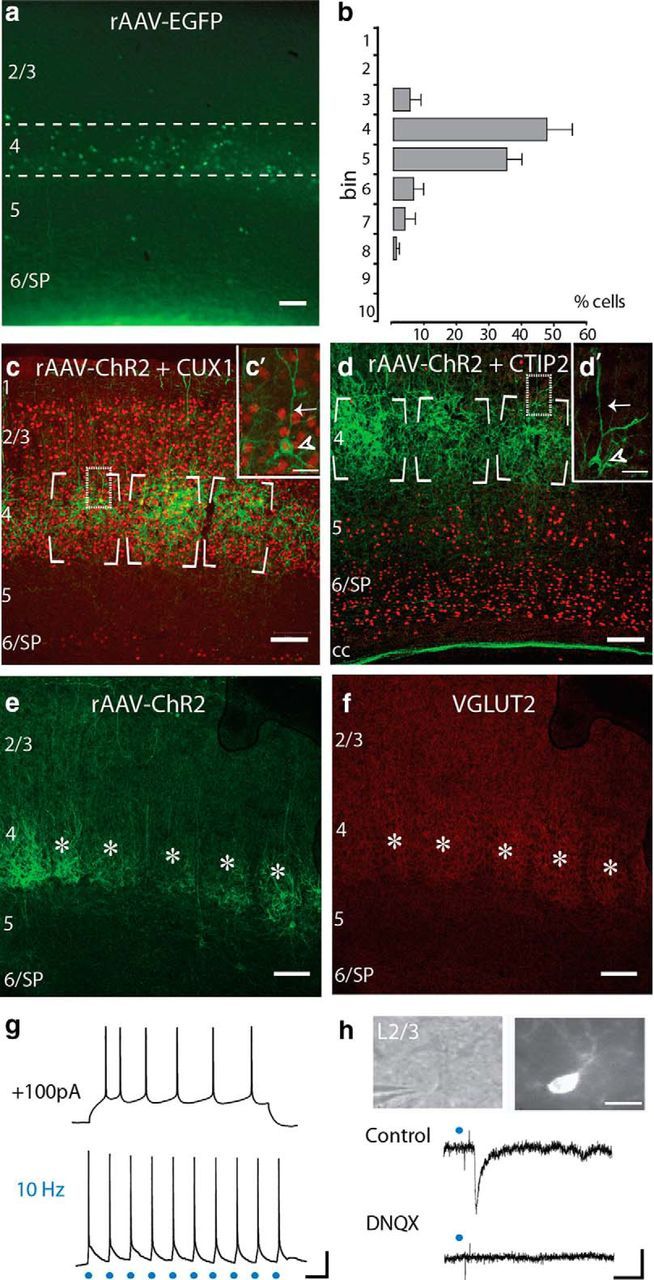
Figure 1.

Ebf2 labels L4 neurons in somatosensory cortex. a, Coronal section through S1 of an adult Ebf2-Cre mouse injected at E15 with rAAV-EGFP. b, Cell quantification of Ebf2+ cells across cortical layers. Dashed lines in a delineate L4 (bins 4–5). Approximately 84% of Ebf2+ cells in the neocortex of Ebf2-Cre;rAAV-EGFP mice were in L4. c, d, Adult coronal Ebf2-Cre;rAAV-ChR2 sections immunostained against Cux1 (c) and Ctip2 (d; both red) to label boundaries of L2–4 and L5–6, respectively. Insets (c′, d′) are confocal sections of representative pyramidal neurons (arrowheads) with a prominent apical dendrite (arrows). e, f, Ebf2-Cre;rAAV-ChR2 expression at P11 broadly overlaps with VGlut2+ expression (red) at individual barrels (asterisks). g, Representative whole-cell patch-clamp recordings in acute slices of an adult Ebf2+p neuron in response to a depolarizing current injection (100 pA; top) and to optogenetic stimulation (3 ms, 10 Hz; bottom). h, Optogenetic stimulation of Ebf2+ L4 neurons (3 ms, single pulse) triggers an EPSP in a L2/3 pyramidal neuron (middle) targeted under DIC optics (top left), and filled intracellularly with AlexaFluor 594 (top right). The response is completely blocked by DNQX (bottom). Scale bars: a–f, 100 μm; c′, d′, 50 μm; h (top), 20 μm; g, h, 20 mV and 100 ms.
