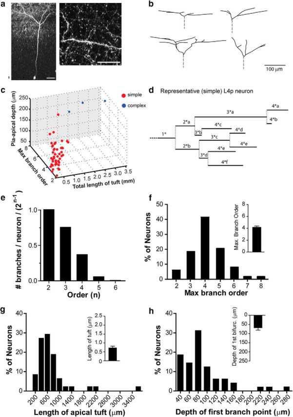
Figure 2.

Morphology of Ebf2+ pyramidal neurons in L4 (apical tufts). a, Two-photon image of a representative Ebf2+ L4 pyramidal neuron (soma depth ∼453 μm) and a dendritic segment in L1 (max proj, 17 slices, 2 μm apart) acquired in vivo in an adult Ebf2-Cre;rAAV-EGFP mouse. Scale bars: a, 50 μm; b, 100 μm. b, Neurolucida reconstructions of apical dendritic tufts from four representative neurons imaged in vivo. c, Ebf2+p neurons were segregated into two groups, simple (red) and complex (blue), using a k means test in MATLAB (see Materials and Methods) using values in f–h. d, Representative dendrogram of the L4 Ebf2+p apical tuft shown in a. e, Fraction of neurons with dendrites of a given order “n” (first order values, primary dendrites are excluded). f–h, Frequency distribution histograms for maximum branch order (f), total length of the apical dendritic tuft (g), and depth of first bifurcation from the pial surface (h) for all reconstructed Ebf2+p neurons (n = 48). Insets show mean ± SEM.
