Fig. 4.
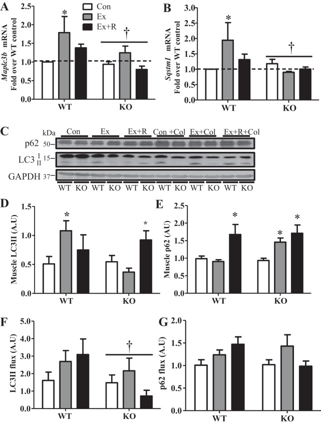
Expression of autophagy genes and induction of autophagy with exercise are differentially regulated in PGC-1α KO animals. A and B: gene expression of autophagy factors, microtubule-associated protein 1 light chain 3 [Maplc3b (LC3)] and sequestosome 1 [Sqstm1 (p62)], in WT and KO animals in Ex and Ex + R groups compared with WT animals in Con group. Gapdh and Actb were used as housekeeping genes. C–E: blots and quantification of autophagic protein in whole muscle extracts from WT and KO animals in Con, Ex, and Ex + R groups treated with vehicle or colchicine (0.4 mg·kg−1·day−1) for 2 days. F and G: autophagy flux as determined by percent change in protein content from colchicine- and vehicle-treated WT and KO animals in Con, Ex, and Ex + R groups. GAPDH was used as loading control. Values are means ± SE; n = 5–9. *P < 0.05, significant effect of exercise. †P < 0.05, significant effect of genotype.

