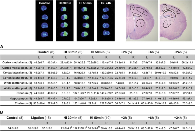
Figure 7.
Regional cerebral blood flow (rCBF) measurements (mL/100 g per minute) by the iodoantipyrine method during and after HI using both calculations from autoradiograms in nine brain regions (A) and liquid scintillation counting (LSC) of whole brain hemispheres (B). The results of the two methodological approaches for measurement of CBF after iodoantipyrine injections were comparable. In addition to results in this figure, the anterior and posterior parts of the medial/lateral and white matter are presented. Small differences were found be tween the anterior/posterior parts in these brain regions although rCBF was almost always somewhat lower in the more posterior parts of medial and lateral cortex. Note also that in animals with left carotid artery ligation only (without HI) CBF at 1 hour after the ligation was near to identical with control animals (see B). Examples of autoradiograms (four levels/animal) are shown above the table where gray scale images have been converted to rainbow color spectrums to more clearly visualize the changes in rCBF. The nine regions for rCBF measurements are shown in top right. Data are mean±s.e.m., number of animal for each group in brackets. Outcome of statistical analysis only presented for data in (B) (see Figure 6 for rCBF). ¤P<0.01, #P<0.001 (ANOVA followed by LSD). L, left (ligated) hemisphere; R, right hemisphere.

