Figure 1.
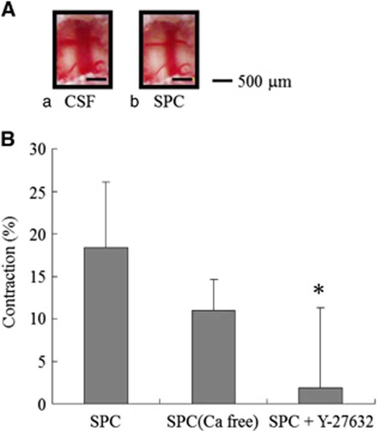
Changes in the rat basilar artery diameter induced by sphingosylphosphorylcholine (SPC). (A) A control rat basilar artery treated with artificial cerebrospinal fluid (CSF) (a) and vasoconstriction with 100 μmol/L SPC (b). Bars, 500 μm. (B) Percent reduction in basilar artery diameter after treatment with 100 μmol/L SPC, 100 μmol/L SPC and Ca2+-free CSF (n=5), and 10 μmol/L Y-27632 (n=9). Values are shown as means+s.d. *P<0.001 versus 100 μmol/L SPC.

