Figure 6.
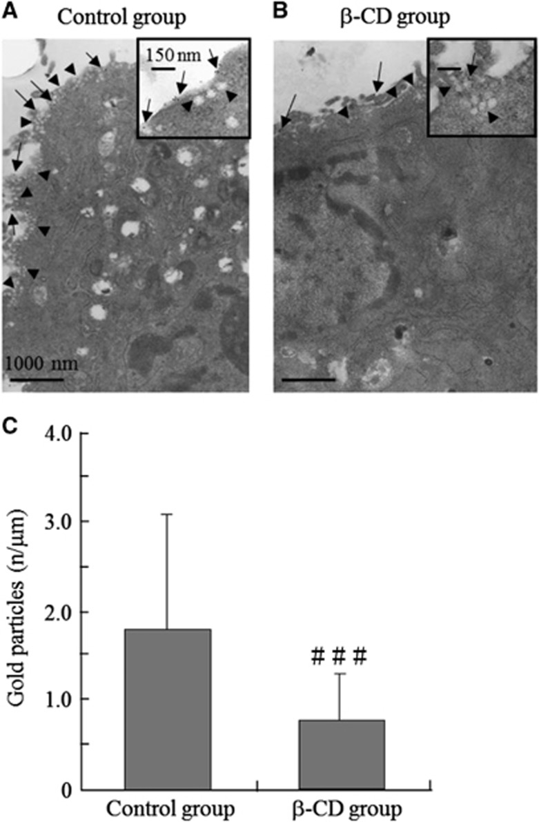
Effect of β-cyclodextrin (β-CD) on formation of raft clusters in vascular smooth muscle (VSM) cells. (A) Labeling of human brain VSM cells (HBVSMCs) with the raft marker ganglioside GM1 (Control group). Bar, 1,000 nm. (B) GM1 labeling of HBVSMCs treated with β-CD (β-CD group). Bar, 1,000 nm. HBVSMCs treated with β-CD showed a marked decrease in the GM1 count (gold particles, arrows) compared with the control group. Arrowheads revealed caveolar structures, defined as uncoated 50- to 100-nm surface invaginations with clear connections to the plasma membrane. The insets in (A) and (B) show higher magnification images (Bars, 150 nm). (C) The GM1 count (gold particles) per unit membrane length (n/μm) was higher in nontreated cells compared with β-CD-treated cells. Values are shown as means+s.d. ###P<0.05 versus control group.

