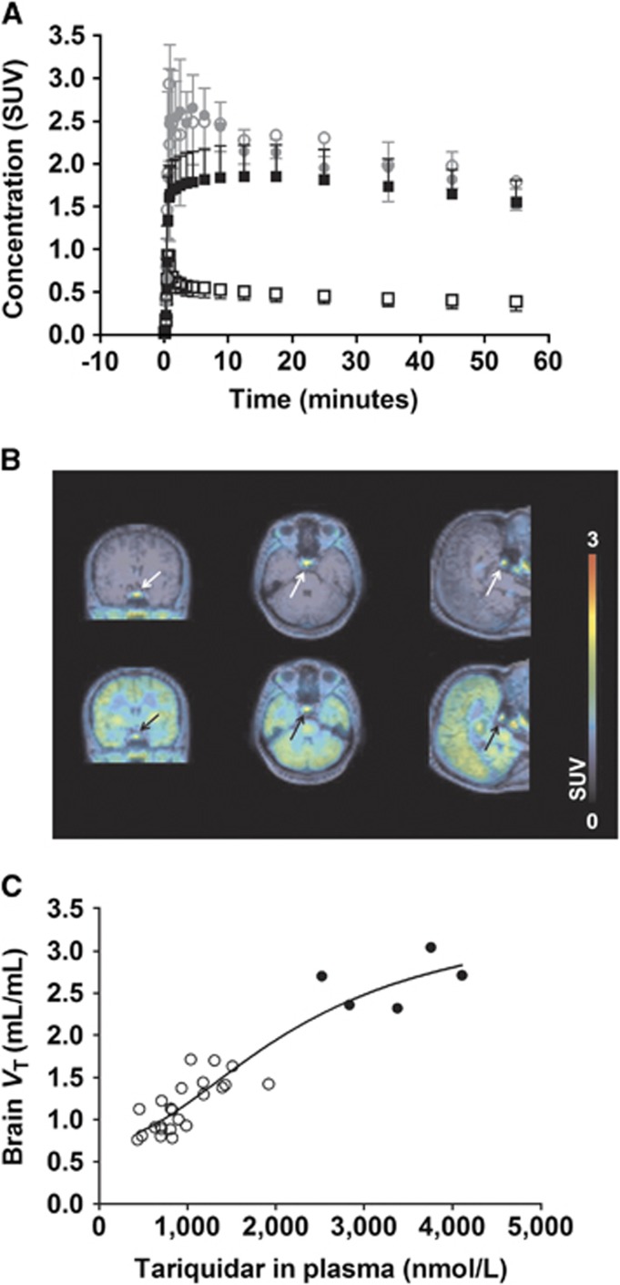
Figure 1.
(A) Time-activity curves (TACs) (mean standardised uptake value, SUV±s.d., N=5) of (R)-[11C]verapamil in whole-brain gray matter (black) and pituitary gland (gray) before (open symbols) and during (filled symbols) tariquidar infusion. (B) Magnetic resonance (MR)-coregistered PET average images (0 to 60 minutes) of (R)-[11C]verapamil before (upper row) and during (lower row) tariquidar infusion in coronal (left), transaxial (middle) and sagittal (right) views. Radioactivity scale is expressed as SUV. The pituitary gland is indicated by an arrow. (C) Extended concentration-effect curve of tariquidar, in which data from this and two previous studies were pooled.7, 14 Data points from this study are shown as filled circles and from previous studies as open circles. The following parameter estimates (mean±standard error) were obtained: half-maximum effect concentration (EC50): 2,248±987 nmol/L; Hill-slope (n) 2.0±1.1; minimum VT: 0.75±0.26; maximum VT: 3.5±1.2; coefficient of determination (R2): 0.88.

