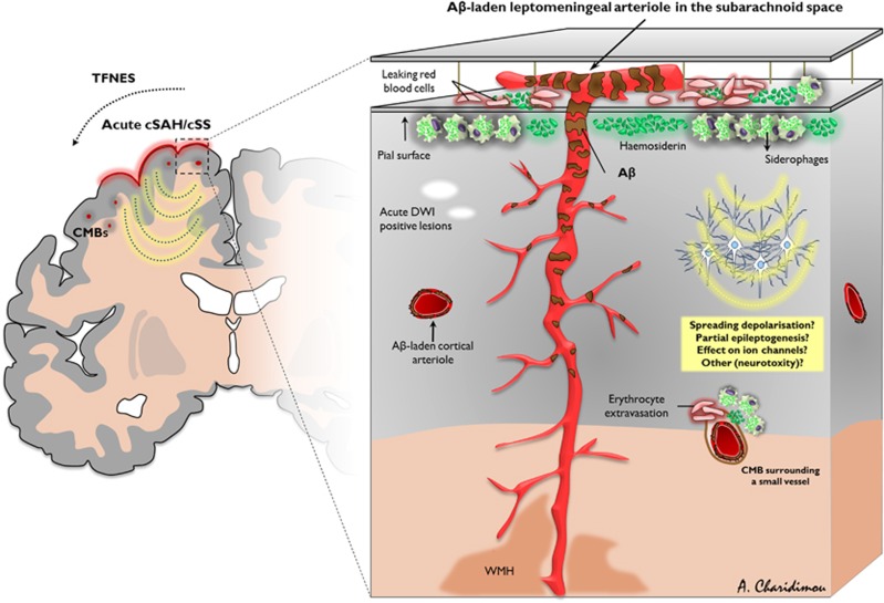
Figure 1.
Schematic diagram of leptomeningeal and superficial parenchymal arterioles showing key features of acute convexity subarachnoid hemorrhage (cSAH) and cortical superficial siderosis (cSS) in sporadic cerebral amyloid angiopathy (CAA). Leptomeningeal and perforating arterioles of a brain section showing amyloid-β (Aβ) deposits (brownish discoloration). Note the reducing vascular Aβ severity moving from the cortical surface into the cerebral white matter. Repeated episodes of hemorrhage (acute cSAH, which can be either clinically silent or symptomatic) from these brittle leptomeningeal or very superficial cortical vessels into the adjacent subarachnoid space are probably an important cause of acute cSAH and cortical superficial siderosis in the chronic phase, which has a characteristic predilection for the cerebral convexities, reflecting linear blood residues, including haemosiderin and hemosiderin-laden macrophages (siderophages) in the subarachnoid space or superficial subpial cortical layers. However, the exact pathophysiologic mechanisms have not yet been proven in detail. Acute cSAH and cSS may induce transient focal neurologic dysfunction (related to spreading depolarization, partial seizure activity focal seizure activity, or other mechanisms), acute microinfarcts on diffusion-weighted (DWI) magnetic resonance imaging (MRI) or be associated with future risk of intracerebral hemorrhage. CMB, cerebral microbleed; TFNEs, transient focal neurological episodes; WMH, white matter hyperintensities.

