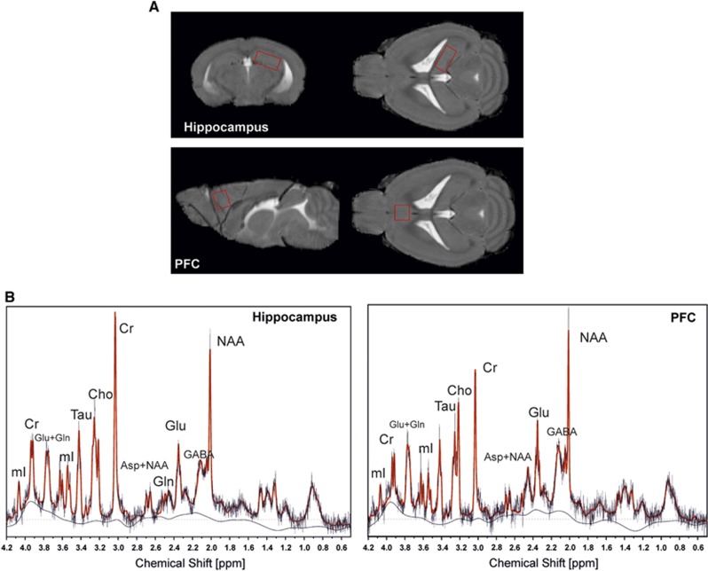
Figure 1.
Voxel location and representative spectra. Voxel localization (A) and typical spectra at 9.4 T (point resolved spectroscopy (PRESS), 3.2 μL, echo time (TE)=10 ms, repetition time (TR)=4 seconds, 256 averages) (B). Both spectra are overlaid by the LC Model fit-curve. Peak resonances are N-acetylaspartate (NAA), creatine and phosphocreatine (Cr), choline-containing compounds (Cho), myo-inositol (mI), glutamate (Glu), glutamine (Gln), Glu+Gln (Glx), taurine (Tau), and γ-aminobutyric acid (GABA).

