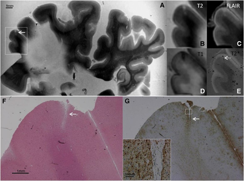
Figure 1.
Intracortical chronic gliotic noncavitated microinfarct (indicated by arrow), identified on magnetic resonance imaging (MRI), in postmortem brain of a 78-year-old female with mild Alzheimer pathology (Braak & Braak (BB) II) and cerebral amyloid angiopathy (CAA). The intracortical gliotic microinfarct appeared hyperintense on T2 (A, enlarged in B) and FLAIR (C), and hypointense on T1 (D). On T2* a vessel (possibly filled with air or clotted blood) could be seen at the same location (E; broken arrow). On Hematoxylin & Eosin (HE) the microinfarct was identified as a region of tissue pallor, accompanied by neuronal death and gliosis (F). The adjacent section was GFAP positive, indicating the presence of astrogliosis (G; inset is enlargement of area indicated with white square). FLAIR, fluid-attenuated inversion recovery; GFAP, glial fibrillary acidic protein.

