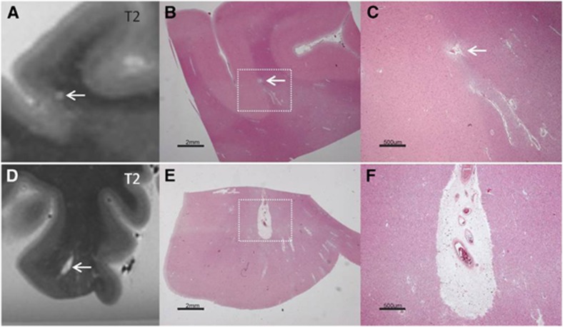
Figure 5.
Two juxtacortical perivascular spaces (PVSs), identified on magnetic resonance imaging (MRI), in postmortem brain of a 78-year-old female with Alzheimer pathology (Braak & Braak (BB) II) and moderate cerebral amyloid angiopathy (CAA) (top row), and in postmortem brain of a 68-year-old female with Alzheimer pathology (BB VI) and severe CAA (bottom row). The first PVS was located on the gray/white matter border and had the same MRI features as chronic gliotic microinfarcts (A). On Hematoxylin & Eosin (HE) there was no evidence of neuronal death or gliosis (B, enlarged in C). The second enlarged-appearing tubular-shaped PVS was located perpendicular to the cortex and had the same MRI features as chronic gliotic microinfarcts with cavitation (D). On HE there was no evidence of neuronal death or gliosis (E, enlarged in F).

