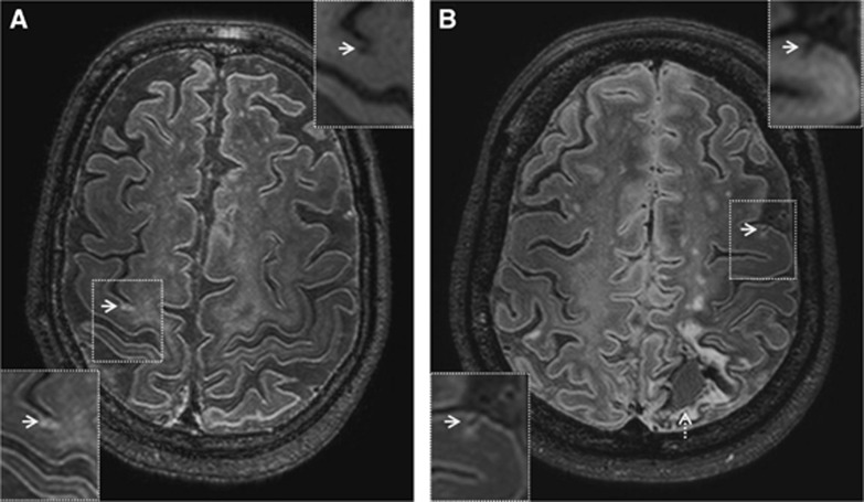
Figure 6.
Cortical microinfarct subtypes on in vivo 7-tesla FLAIR (0.8 mm isotropic voxels) magnetic resonance imaging (MRI). A chronic gliotic microinfarct in a 76-year-old healthy male (A). A chronic gliotic microinfarct with cavitation in a 42-year-old female with spontaneous intracerebral lobar hemorrhage (indicated by broken arrow) (B; courtesy of Dr CJM Klijn, UMCU, The Netherlands). Insets in upper right corner are T1-weighted images (1.0 mm isotropic voxels). Chronic gliotic microinfarcts with and without cavitation are both hypointense on in vivo T1-weighted images. FLAIR, fluid-attenuated inversion recovery.

