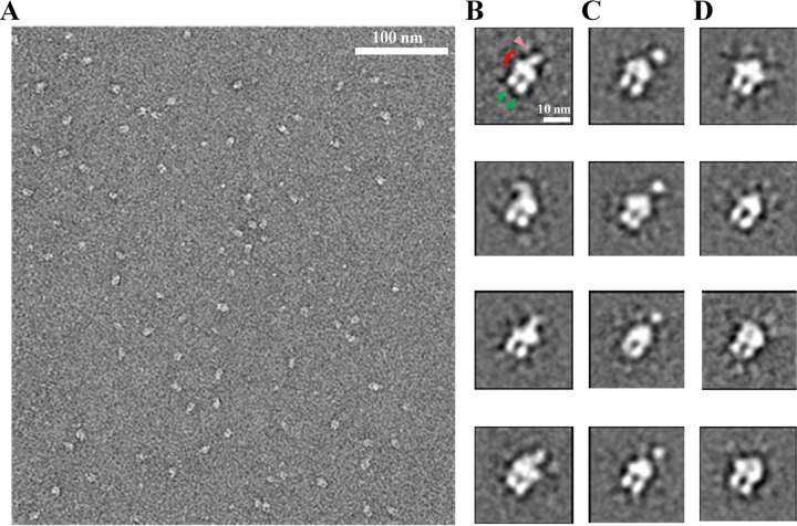
FIG 4.
2D image analysis of the ExbB4-ExbD1-TonB1 complex. (A) A typical TEM micrograph showing uniform particles of ∼10-nm diameter in various orientations. Following extraction, single particles were classified using ISAC. (B) Four class averages showing the DDM micelle (bracket), the cytoplasmic domains of ExbB (green arrowheads), and dimerized protein in the periplasmic domain (pink arrowhead). These averages show dimerization beginning at the micelle, whereas the periplasmic dimerization in panel C is at a point further from the micelle. (D) Four class averages with no observable density on the periplasmic side of the micelle. Single, extended polypeptides are not expected to be visualized by negative-staining EM.

