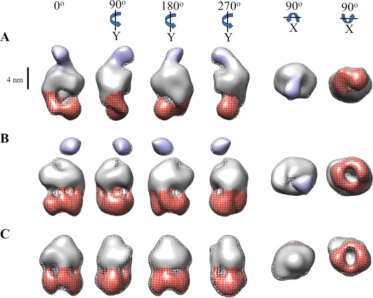
FIG 5.
3D volumes representing the consensus conformational states of the ExbB4-ExbD1-TonB1 complex. Following 3D classification using RCT reference models, particles were grouped and used for refinement. The resultant volumes represent consensus structures of the ExbB4-ExbD1-TonB1 complex in three conformational states: extensive periplasmic dimerization (A), distal periplasmic dimerization (B), and no observed dimerization on the periplasmic side of the micelle (C). Volumes in panels B and C show four regions of density descending from the micelle, which can be attributed to a tetramer of ExbB. The distal portions of these cytoplasmic domains form a ringlike arrangement below and parallel to the micelle. The volume in panel A shows two thick regions of density descending unequally from the micelle, attributable to a dimer of ExbB dimers. All rotations are relative to 0°. The mesh contour represents volumes of ∼230 kDa, with the inner volume threshold decreased to display features. A scale bar (4 nm) delineates the potential CM boundary. Cytoplasmic and periplasmic domains are colored red and purple, respectively.

