FIG 2.
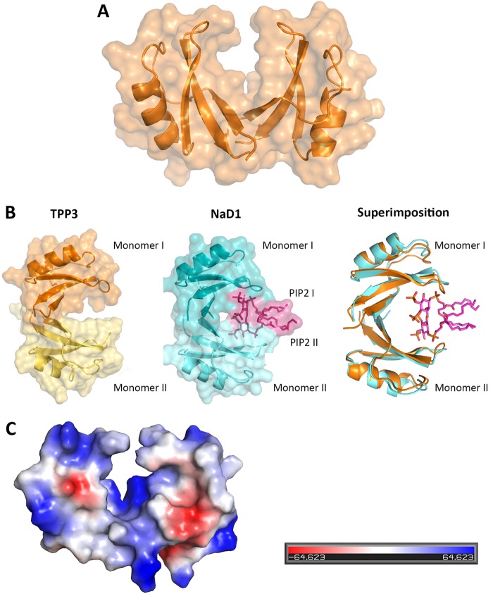
Crystal structure of TPP3. (A) Cartoon representation of the TPP3 structure (orange) shown as a dimer in the asymmetric unit with the molecular surface shown in pale orange. (B) Surface representations of TPP3 and NaD1, displaying the “cationic grip” formed between the two TPP3 monomers (orange and pale yellow), the NaD1 cationic grip (teal and aqua) in which two PIP2 molecules are present (magenta), and the superimposition of the TPP3 dimer (orange) over the NaD1 dimer containing PIP2 molecules (aqua and magenta). (C) Qualitative electrostatic surface representation of TPP3 (blue is positive, red is negative, and white is uncharged or hydrophobic). This figure was generated using Pymol.

