FIG 5.
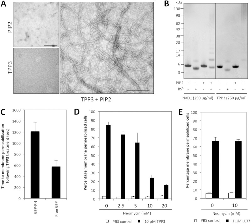
TPP3 interacts with PIP2. (A) Negative-staining TEM images of TPP3 showing the formation of fibrils in the presence of PIP2. Scale bars represent 200 nm. (B) Cross-linking analysis of TPP3 with PIP2. The ability of TPP3 to form oligomers in the presence of PIP2 was investigated using the amine reactive cross-linker BS3, followed by SDS-PAGE and Coomassie staining. (C) Ability of TPP3 to permeabilize tumor cells in GFP-PH-overexpressing cells. HeLa cells overexpressing either GFP-PH or free GFP were treated with TPP3 and imaged with CLSM to determine the mean time to membrane permeabilization. Statistical analysis was performed using an unpaired, two-tailed t test (n = 26 for GFP-PH and n = 21 for free GFP-expressing cells; P = 0.001). (D) Ability of neomycin to inhibit TPP3-mediated U937 cell lysis in a concentration-dependent manner, as determined by flow cytometry. (E) Ability of neomycin to inhibit LL-37-mediated U937 cell lysis. For panels D and E, error bars represent SEMs (n = 3); results are representative of those from three independent experiments.

