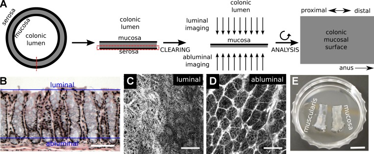
Fig. 1.
Deep mucosal imaging (DMI) is a workflow for imaging and reconstructing extended volumes of colorectal mucosa. A: the mouse colon is opened, and the mucosal layer is isolated, cleared, and imaged in a whole mount setup. B–D: the conventional plane of analysis in hematoxylin and eosin (H&E)-stained histological sections (B) is tangential to the luminal (C) and abluminal (D) planes in whole mount imaging of colons from mTmG mice (C and D). E: separation of the myenteric layer was essential for clearing in the distal colon and rectum. Scale bars: 50 µm (B), 75 µm (C and D), and 8.75 mm (E).

