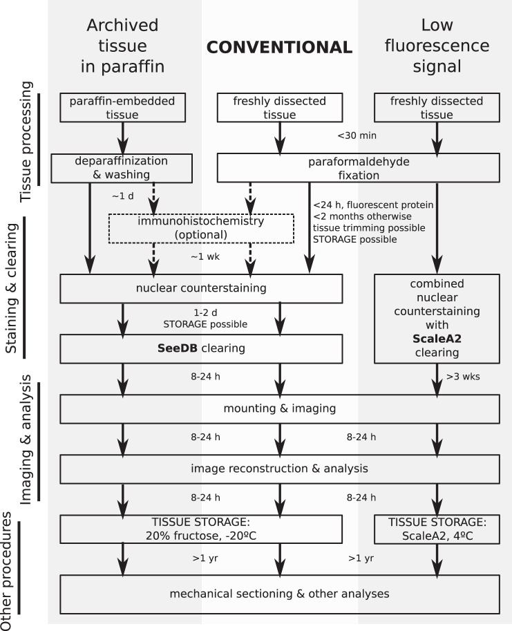
Fig. 10.
Schematic of the DMI workflow. Most applications using freshly dissected mouse or human colonic tissue should follow the conventional workflow (center), which utilizes SeeDB as the clearing agent. However, if the expression of fluorescent proteins in the sample is low, ScaleA2 should be used as the clearing agent (right). Recovery and imaging of paraffin-embedded tissue involves deparaffinization and clearing with SeeDB (left). The arrows between steps indicate the amount of time needed by the previous step. Tissue can also be stored in 20% fructose or ScaleA2 after completion of certain steps, as shown on the diagram, for synchronous processing of experimental samples.

