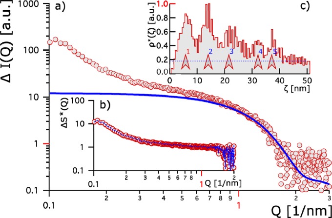
FIG 3.

(a) Red circles show the relative change in the scattering contrast of Thc_Cut1 plus HFB4 from that of Thc_Cut1 plus HFB9. The bold blue line represents the hypothetical Thc_Cut1 form factor. (b) Changes in the apparent structure factor between the two systems. Red circles indicate the apparent structure factor, and the blue line gives the fit thereto. (c) The apparent pair correlation [ρ*(ζ)] is given as a function of the relative distance ζ. It is normalized to 1. Five peaks (marked by red arrows) are identified and give evidence for stronger clustering of Thc_Cut1 plus HFB9b than of Thc_Cut1 plus HFB4. The first peak (1 [red]) evolves at a distance equivalent to a protein size of 5 nm; blue-numbered peaks 2 to 5 indicate longer-range interactions and hint of stronger aggregation.
